


Abstract
Some specific features of the transport of silver nanoparticles onto a SiO2 substrate under focused nanosecond IR laser pulses is experimentally investigated. A possibility of obtaining silver coatings is demonstrated. The formation of silver nanostructures as a result of pulsed laser ablation in air is studied. Nanoparticles are formed by exposing a silver film to radiation of an HTF MARK (Bulat) laser marker (λ = 1064 nm). The thus prepared nanoparticles are analysed using scanning electron microscopy and optical spectroscopy.
Export citation and abstract BibTeX RIS
| A.A. Nastulyavichus, N.A. Smirnov, A.A. Ionin, I.N. Saraeva, N.I. Busleev, A.A. Rudenko, R.A. Khmelnitskii, D.A. Zayarnyi P.N. Lebedev Physical Institute, Russian Academy of Sciences, Leninsky prosp. 53, 119991 Moscow, Russia |
| e-mail: ganuary_moon@mail.ru |
| S.I. Kudryashov P.N. Lebedev Physical Institute, Russian Academy of Sciences, Leninsky prosp. 53, 119991 Moscow, Russia; ITMO University, Kornverkskii prosp. 49, 197101 St. Petersburg, Russia |
1. Introduction
Many works devoted to the preparation of metal (in particular, silver) nanoparticles have been published recently. The interest in these objects is due to the prospects of their application in different fields of science and technology, including the use in nanophotonics and nanoplasmonics devices, development of biosensors, and improvement of the solar cell efficiency [1 – 3]. Using nanoparticles, one can control micro- and macro-processes (for example, interaction of laser radiation with matter) and observe the position of local surface plasmon resonance [4, 5]. Varying the main parameters of nanoparticles (shape and sizes), one can control the position of the optical absorption peak for structures based on them. The presence of structural inhomogeneities, including surface roughness and gaps between particles, may enhance the effects of Raman scattering and optical nonlinearity due to the high intensity of plasmon electromagnetic fields [6].
Nanoparticles can be obtained in different ways: lithographic [7] and chemical [8] methods, precipitation of colloidal solutions [9], and deposition in vacuum and gases [10]. All these techniques have their own advantages and drawbacks. In this study, we applied laser transport of silver nanoparticles (formed from thin magnetron-sputtered silver films) onto SiO2 substrates. This method can be used to form nanostructures. It is nontoxic and allows nanostructures with specified characteristics to be formed. Its other advantages are purity and stability of particles formed [11, 12].
2. Experimental
Silver films were prepared by magnetron sputtering in argon on SiO2 substrates. Then these films were exposed to radiation of an HTF MARK fibre laser marker (Bulat) on Yb3+ ions with a laser pulse FWHM of 120 ns, maximum pulse energy of 1 mJ, and pulse repetition rate up to 80 kHz (Fig. 1a). The laser beam was focused by a galvano-scanner with an objective focal length of 160 mm. Transmission spectra of the samples were recorded on a V-70 spectrometer (Bruker, Germany). The size distribution of silver particles was analysed by visualising samples with a JEOL 7001F scanning electron microscope (SEM) (Japan); before the visualisation, a thin copper film was deposited on the samples to exclude surface charging. The chemical composition of the silver structures obtained was confirmed by energy-dispersive X-ray spectroscopic chemical microanalysis using the INCA module (Oxford Instruments, England) of the electron microscope.

Figure 1. (a) Photograph of the experimental setup [HTF MARK laser marker (Bulat)] and (b) schematic diagram of laser ablation.
Download figure:
Standard image3. Results
The experiment was performed on silver films with thicknesses of 20, 30, 45, 60, and 90 nm. An area 5 × 5 mm in size was chosen on the surface of each film, and an object glass was installed above the films. The transport of silver particles onto the glass substrate was performed by ablation in air under multipulse irradiation of silver films by a focused laser beam. A schematic of this process is shown in Fig. 1b. Scanning was carried out at the maximum pulse energy at which the plasma plume formed near the target surface did not structurize the glass onto which the particles were transported. The energy density F0 on the film was 15 J cm−2 in pulse at a rate of 40 kHz and a scan rate of 5 mm s−1. The minimum size D0 of the focusing spot was 26 μm. A melt of nanoparticles formed as a result of ablation leaved the target to fall on the glass substrate, where clusters of nanoparticles were finally formed. The morphology of the thus obtained structures is presented in Fig. 2. The particle diameter d lies in the range of 10 – 80 nm. The average particle diameter for different films varies from 17 to 46 nm.
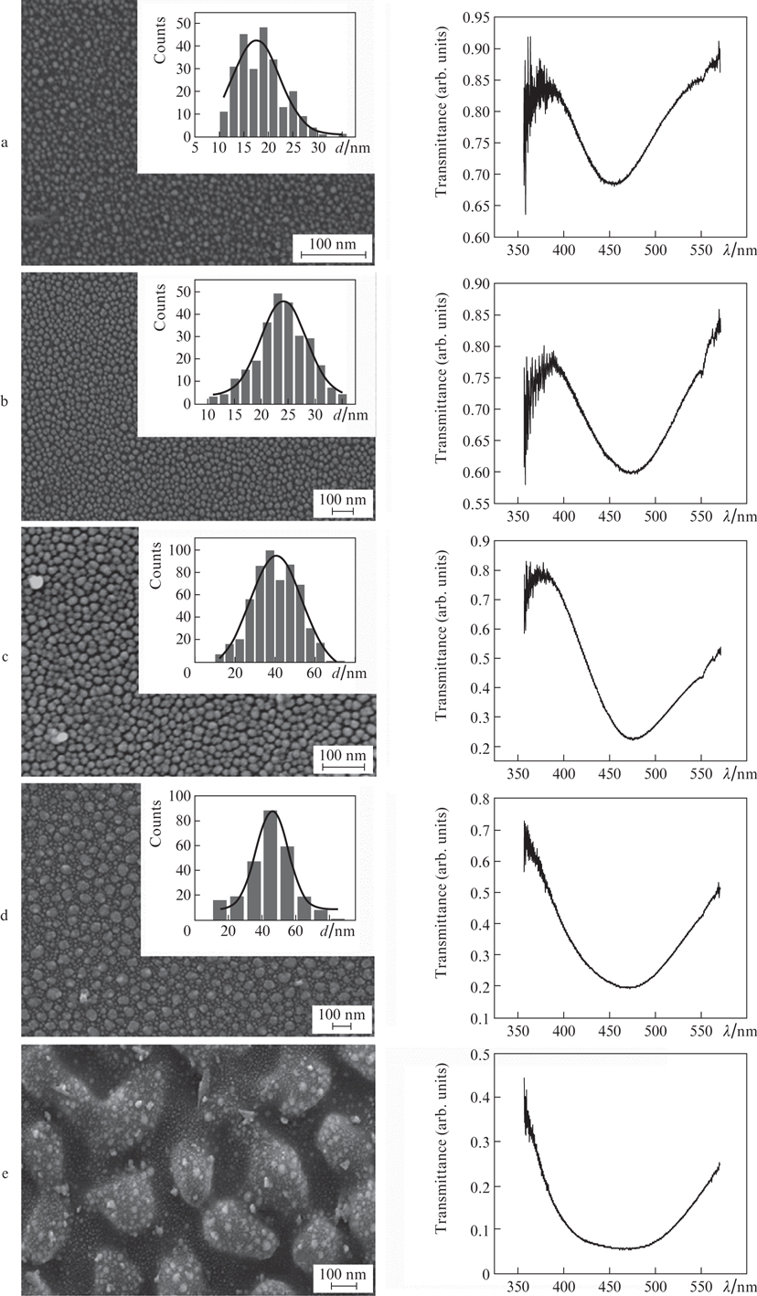
Figure 2. SEM images of structures, particle-size distributions, and optical transmission spectra of samples with silver nanoparticles for film thicknesses of (a) 20, (b) 30), (c) 45, (d) 60, and (e) 90 nm.
Download figure:
Standard image4. Discussion
The SEM images in Fig. 2 show that the coatings obtained from films with thicknesses of 20, 30, 45, and 60 nm consist of arrays of individual silver nanoparticles, whose shape is close to spherical and maximum diameter is ∼100 nm. In the case of 90-nm-thick film, individual nanoparticles merge with each other (because their number is large), as a result of which there is a significant spread in their size. Note that, due to the large amount of ejected material, a thin film with embedded alloyed particles of larger size (nanostructured film) is formed on the sample surface rather than a monolayer of particles. This surface structure does not allow one to construct a histogram of particle-size distribution. The plasmon resonance peak in the transmission spectra lies in the vicinity of λ ≈ 450 nm; with an increase in the thickness of the film used for ablation, this peak becomes red-shifted and broadened, which is caused by an increase in the size of formed nanoparticles and their larger spread in size.
Since the experiment was performed in air, oxide could be formed on the particle surface, as evidenced by the plasmon resonance red shift [13]. To verify this suggestion, we performed a numerical simulation by the FDTD (finite-difference time-domain) method. It was assumed that a plane electromagnetic linearly polarised wave is incident on a nanoparticle. Then absorption cross sections were calculated for different wavelengths. Using certain boundary conditions, we took into account the influence of neighbouring particles, which were spaced at a distance of 10 nm. Three cases were considered: (i) a silver particle 20 nm in diameter, (ii) a particle coated by a 5-nm-thick oxide shell, and (iii) a particle coated by a 10-nm shell. The simulation results show that the presence of a 5-nm shell is sufficient for the red shift of the maximum of absorption cross section up to λ ≈ 450 nm; this conclusion is consistent with the experimental data (Fig. 3). In addition, there may be a contribution from the hybridisation of silver oxide atomic orbitals, which affects the plasmon resonance peak splitting. Numerical calculations showed also that the substrate does not affect the peak shift.
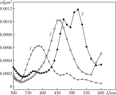
Figure 3. Dependences of the absorption cross section σ on wavelength λ for (1) silver particles with d = 20 nm and (2, 3) the same particles coated by oxide layers with thicknesses of (2) 5 and (3) 10 nm.
Download figure:
Standard imageThe structures formed by ablation of thin films contain small particles. Their size is explained by the limited amount of silver initially present in the film and then deposited on the glass. An analysis of the experimental data showed that the size of silver nanoparticles linearly depends on the evaporated film thickness (Fig. 4).

Figure 4. Dependence of the average diameter dav of particles in the nanostructures on the thickness of the film subjected to ablation and its linear approximation.
Download figure:
Standard image5. Conclusions
Silver nanoparticles were obtained in experiments with irradiation of thin silver films by nanosecond IR laser pulses (λ = 1064 nm); these particles were deposited by ablation of films in air onto a glass substrates. An increase in the film thickness led to a linear growth of particles in size, due to which their transmission spectra became broadened and red-shifted. A further increase in the film thickness led to merging of particles because of the large amount of material transported from the film surface to the substrate.
Acknowledgements
This study was performed within State Contract No. 0023-20160002.